History and Overview
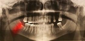
Everyone knows what a cavity is, but cavitations are much less well known. Both words come from the same root word, “hole.” A cavity is a hole in the tooth, whereas, a cavitation is a hole in the bone that cannot be detected through visual inspection.

The term “cavitation” was coined in 1930 by a well-known orthopedic researcher to describe a disease process in which the lack of blood supply to an area of bone resulted in a hole or “hollowed out” portion of the jawbone or other bones in the body.
As we mentioned last time, G. V. Black also described this process in 1915 as a progressive disease of the jawbone that kills bone cells and produces large hollowed out areas of bony tissue or a soft mass enclosing particles of necrotic (dead) bone. He was intrigued by the unique ability of this disease to produce extensive jawbone destruction without causing redness, swelling of the overlying film or increasing the patient’s body temperature. The progressive impairment of blood supply to the marrow of the bone essentially produces small “heart attacks” (infarcts) in the jawbone, thus resulting in osteonecrosis (bone death). It is possible that a biofilm form of the bacteria, which is antibiotic resistant, adheres to the inside of the capillary walls, and its toxins and by-products, combining with other cellular material, are responsible for the infarct producing clots. Black suggested that surgically removing this dead necrotic tissue was necessary to promote healing of the jawbone.
Current Use of the Term “Cavitation”
In the last decade, the term “cavitation” has been used not only to describe lesions appearing as empty holes but also various types of lesions in the jawbone found through tissue analysis to be ischemic (lacking in oxygen), necrotic (dead), osteomyelitic (bone infected) and toxic. These lesions are often located in old extraction sites and under or near the roots of root canal teeth, cavital (dead) teeth and wisdom teeth. Sometimes, they seem to spread extensively from these locations throughout the jawbone and may penetrate the sinuses or totally encompass the inferior alveolar (jaw) nerve.
Recent Research
Recent research by Dr. Boyd Haley shows ALL cavitation tissue samples tested contain toxins, which significantly inhibit one or more of five basic body enzymes necessary in the energy production cycle. These small chemical toxins, metabolic waste products (most likely from anaerobic bacteria) may produce significant systemic effects, as well as play an important role in the localized disease process, which negatively affects the blood supply in the jawbone. There are indications that when these toxins combine with chemicals or heavy metals such as mercury, more potent toxins may be formed.
Research from Germany indicates the jawbone may be a holding tank for chemicals and heavy metals (especially wisdom teeth sites).
Clinical experience indicates it is sometimes difficult for some patients to successfully detoxify mercury from the body until after cavitations, as well as fillings containing mercury are removed.
NICO – Cavitations Accompanied by Pain
The term NICO, which stands for “neuralgia-inducing cavitational osteonecrosis,” has been used when severe facial pain, neuralgia, headache or a phantom toothache accompanies this disease. Although the presence of cavitations is a common occurrence, only a small percentage of the individuals with cavitations suffer from the pain component included in the description of NICO lesions. Even if pain symptoms or localized jawbone symptoms are not present, systemic symptoms can be extensive. The intense concern expressed by several researchers and physicians earlier this century about the systemic influences of these lesions has likewise become a concern for contemporary dentists, physicians, and researchers.
Occurrence of Cavitations – CAVITAT
Bob Jones, inventor of the CAVITAT (an ultrasound instrument designed to detect and image cavitations), reported finding cavitations of various sizes and severity in approximately 94% of several thousand wisdom teeth sites he scanned. He also reported finding cavitations under or located near 100% of root canal teeth scanned in both males and females of various ages from several geographic areas of the United States.
Initiating – Predisposing – Risk Factors
There are several possible initiating, predisposing and risk factors associated with cavitations. It is likely that a combination of these factors present in a particular individual in a particular jawbone area will influence the occurrence, type, size, progression and growth pattern of a lesion. One of the major initiating factors is likely dental trauma, which includes physical, bacterial and toxic components.
- Physical Trauma
- Extractions
- Dental injections
- Periodontal surgery
- Root canal procedures
- Grinding – bruxism
- Electrical trauma/metallic restorations
- High speed drilling
- Bacterial trauma
- Periodontal disease
- Cysts
- Abscesses
- Root canal bacteria
- Incomplete clean out after extractions
- Infected wisdom teeth or tooth buds
- Toxic trauma
- Dental materials
- Root canal toxins
- Anesthetic by-products
- Vasoconstrictors in anesthetics
- Chemical toxins
- Bacterial toxins
- Other toxins
Predisposing Factors
Predisposing factors include clotting disorders such as thrombophilia and hypofibrinolysis (which may be undiagnosed); age (evidence suggests that as many as 11% of older individuals may have major or complete blockage of arteries feeding the jaws); radiation or chemotherapy for cancer; rheumatoid arthritis, lymphoma or bone dysplasia; variable atmospheric pressures in occupation, osteoporosis, lupus, sickle cell disease, homocystinemia or Gaucher’s disease; hyperlipidemia, hemodialysis, gout or antiphospholipid antibody syndrome; inactivity (bedridden, paraplegic); and deficiency of thyroid or growth hormone. Risk factors that may be responsible for schemic osteonecrosis include corticosteroid use, pregnancy, estrogen use, alcoholism, cigarette smoking and pancreatitis.
Wisdom Teeth Sites
One source of data indicates that 45% of all jawbone cavitations are located in the third molar (wisdom teeth sites). These areas are particularly predisposed because they contain small terminal vessels (microvasculature), and osteonecrosis is a disease of such vessels. Injections for dental procedures are often given near these areas. If the local anesthetic used contains a vasoconstrictor (often epinephrine), it may shut down the blood supply to the bone in these areas. For this reason, the use of non-vasoconstricting anesthetics is indicated.
Recommended Treatment
The recommended treatment of cavitations at the present time remains the same as that proposed by G. V. Black: surgical debridement (scraping clean) of the area to remove all unhealthy bone and all pathology such as abscesses and cysts. It is not sufficient to “punch” a small hole in the bone, drill a little and rinse it out. In fact, this, and the practice of injecting these lesions with homeopathics and other substances may very well increase the severity of the lesion instead of lessening it.
After the unhealthy bone is removed, the goal is bone regeneration. Up to this point in time, successful bone regeneration has relied a great deal on the healing capacity of the individual’s body and the treatment or elimination of predisposing and risk factors, which is not always possible. Lack of healing or reoccurrence of a lesion and the need for retreatment is always a possibility, no matter how well the surgery is performed. There are very few dentists who are trained in effectively diagnosing and treating these lesions. Those who are not so trained are not qualified to diagnose this condition or confidently assure patients that they do not have cavitations.
Prevention of Cavitations
Prevention of cavitations involves the elimination or appropriate modification of initiating, predisposing and risk factors. There are new instruments, products and technological applications which may improve prevention and treatment procedures and enhance the bone regeneration process. Many questions are yet to be answered, and more research is needed to perfect the prevention, diagnosis and treatment of cavitations, but our knowledge is increasing daily.
Most importantly, many individuals are receiving relief from local and systemic symptoms, diseases and pain by the surgical treatment of cavitations.